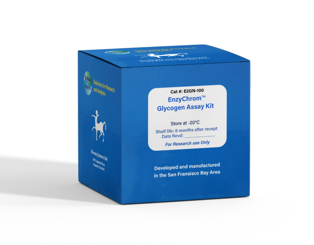
DESCRIPTION
GLYCOGEN is a branched polysaccharide of glucose units linked by α1,4 glycosidic bonds and α-1,6 glycosidic bonds. It is stored primarily in the liver and muscle, and forms an energy reserve that can be quickly mobilized to meet a sudden need for glucose. The most common glycogen metabolism disorder is found in diabetes, in which, due to abnormal amounts of insulin, liver glycogen can be abnormally accumulated or depleted. Genetic glycogen storage diseases have been associated with various inborn errors of metabolism caused by deficiencies of enzymes necessary for glycogen synthesis or breakdown.
Simple, direct and automation-ready procedures for measuring glycogen concentrations find wide applications in research and drug discovery. BioAssay Systems' glycogen assay uses a single Working Reagent that combines the enzymatic break down of glycogen and the detection of glucose in one step. The color intensity of the reaction product at 570nm or fluorescence intensity at λex/em = 530/585 nm is directly proportional to the glycogen concentration in the sample. This simple convenient assay is carried out at room temperature and takes only 30 min.
KEY FEATURES
Use as little as 10 µL samples. Linear detection range: 2 to 200 µg/mL glycogen for colorimetric assays and 0.2 to 20 µg/mL for fluorimetric assays.
KIT CONTENTS (100 TESTS IN 96-WELL PLATES)
Assay Buffer: 12 mL Dye Reagent: 120 µL
Enzyme A: Dried Standard: 50 µL 50 mg/mL
Enzyme B: 120 µL
Storage conditions. The kit is shipped on ice. Store all components at -20°C upon receiving. Shelf life: 6 months after receipt.
Precautions: reagents are for research use only. Normal precautions for laboratory reagents should be exercised while using the reagents. Please refer to Material Safety Data Sheet for detailed information
PROCEDURES
Reagent Preparation: Reconstitute Enzyme A by adding 120 µL Assay Buffer to the Enzyme A tube. Make sure Enzyme A is fully dissolved by pipetting up and down. Store reconstituted Enzyme A at -20°C and use within 1 month.
Sample Preparation:
Samples can be prepared according to Murat & Serfaty (Clin Chem. 20:1576-1577, 1974). Briefly, homogenize tissue/cell sample in 25 mM citrate, pH 4.2, 2.5 g/L NaF on ice. Centrifuge 14,000 g for 5 min to remove debris, and use 10 µL clear supernatant for the assay.
Colorimetric Procedure:
1. Equilibrate all components to room temperature. During experiment, keep thawed enzymes in a refrigerator or on ice.
2. Standards and samples: Dilute standard by mixing 5 µL Standard with 1.245 mL dH2O to give 200 µg/mL standard. Dilute standard in dH2O as follows.
Transfer 10 µL standard and samples into separate wells of a clear flat-bottom microplate. If the sample contains glucose, transfer an additional 10 µL sample to another well for the Sample Blank.
3. Working Reagent. For each reaction well, mix 90 µL Assay Buffer, 1 µL Enzyme A, 1 µL Enzyme B and 1 µL Dye Reagent. For Sample Blank wells, prepare Blank Working Reagent by mixing 90 µL Assay Buffer, 1 µL Enzyme B, and 1 µL Dye Reagent (No Enzyme A). Transfer 90 µL Working Reagent into each Standard and Sample well. Transfer 90 µL Blank Working Reagent to each Sample Blank well. Tap plate to mix.
4. Incubate 30 min at room temperature. Read optical density at 570 nm (550-585 nm).
Fluorimetric Procedure:
For fluorimetric assays, the linear detection range is 0.2 to 20 µg/mL glycogen. Follow steps 1-3 of the colorimetric procedure, but prepare 0, 5, 10, 15 and 20 µg/mL Standard and use a black flat-bottom microplate. Incubate 30 min at room temperature and read fluorescence at λex = 530 nm and λem = 585 nm.
CALCULATION
Subtract Blank reading (OD570nm or fluorescence intensity) from the standard reading values and plot the ∆OD or ∆F against standard concentrations. Determine the slope and calculate the glycogen concentration of the sample.
RSAMPLE and RBLANK are the OD570nm or fluorescence intensity values of the sample and blank (water, or sample blank).
GENERAL CONSIDERATIONS
1. This assay is based on a kinetic reaction, the use of a multi-channel pipettor for adding the working reagent is recommended.
2. SH-group containing reagents (e.g., DTT, β-mercaptoethanol) may interfere with this assay and should be avoided in sample preparation.
MATERIALS REQUIRED, BUT NOT PROVIDED
Pipetting devices, centrifuge tubes, clear flat-bottom uncoated 96-well plates (e.g. VWR cat# 82050-760), optical density plate reader; black flatbottom uncoated 96-well plates (e.g. VWR cat# 82050-676), fluorescence plate reader.
PUBLICATIONS
1. Hunter, A. L., et al (2020). Nuclear receptor REVERBalpha is a statedependent regulator of liver energy metabolism. Proceedings of the National Academy of Sciences, 117(41), 25869-25879.
. Primassin, S., et al (2011). Hepatic and muscular effects of different dietary fat content in VLCAD deficient mice. Mol Genet Metab 104(4): 546-551.
3. Yin, M et al (2011). Metformin improves cardiac function in a
nondiabetic rat model of post-MI heart failure. Am J Physiol Heart Circ
Physiol 301(2):H459-468.
