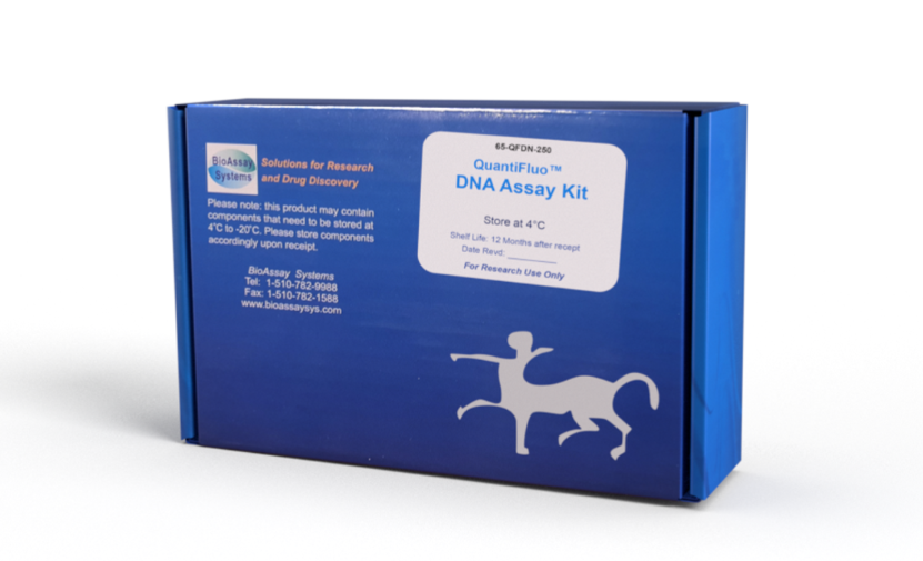
DESCRIPTION
DNA quantitation is a common practice in molecular biology. Very often DNA is available in minute quantities and the traditional UV 260 nm absorbance method requires microgram quantities for reliable results. Accurate determination of DNA concentration, especially when DNA is present at low concentrations, is crucial for reproducible results in sequencing, cloning, transfection and DNA labeling.
Simple, direct and automation-ready procedures for measuring DNA concentration are very desirable. BioAssay Systems' QuantiFluoTM DNA assay kit is designed to accurately measure nanogram quantities of plasmid DNA, cDNA, DNA following polymerase chain reaction and DNA eluted from gels. The improved method utilizes Hoechst dye that binds specifically with double-stranded DNA. The fluorescence intensity, measured at 450nm (exc = 350nm), is directly proportional to the DNA concentration in the sample. The optimized formulation substantially reduces interference by substances in the raw samples.
KEY FEATURES
Sensitive and accurate. Linear detection range 2 ng to 40 ng (100 – 2,000 ng/mL) calf thymus DNA in 96-well plate assay.
Simple and high-throughput. The “mix-and-read” procedure involves addition of a single working reagent and reading the fluorescence intensity. Can be readily automated as a high-throughput assay for thousands of samples per day.
Low interference. RNA, salt (up to 3M NaCl), detergent (< 0.01% SDS) and common DNA extraction buffer do not interfere in the assay.
APPLICATIONS:
Direct Assays: plasmid DNA, genomic DNA, cDNA, DNA following polymerase chain reaction, and DNA extracted from gel and other matrices.
KIT CONTENTS (250 tests in 96-well plates)
Reagent: 50 mL Standard: 1 mL 10 g/mL calf thymus DNA
Storage conditions. The kit is shipped at room temperature. Store the DNA standard at -20°C and the Reagent at 2-8°C. Shelf life: 12 months after receipt.
Precautions: reagents are for research use only. Normal precautions for laboratory reagents should be exercised while using the reagents. Please refer to Material Safety Data Sheet for detailed information.
PROCEDURES
Preparation: bring reagents to room temperature before use.
Procedure using 96-well plate:
1. Prepare 400 L 2000 ng/mL Premix by mixing 80 L Standard and 320 L TE buffer (10mM Tris, 1mM EDTA, pH 7.4). Dilute standards as follows. Transfer 20 L diluted standards and samples into wells of a black flat-bottom 96-well plate. Store standards at 4°C for future use.
2. Add 200 L working reagent and tap lightly to mix.
3. Incubate 1 min at room temperature and read fluorescence emission at 440 - 460nm (peak 450nm, excitation at 340-370 nm).
Procedure using cuvette:
1. Transfer 100 L diluted standards and samples to cuvets.
2. Add 1000 L working reagent and tap lightly to mix.
3. Incubate 1 min at room temperature and read fluorescence intensity at 440 - 460nm (peak 450nm, excitation at 340-360 nm).
CALCULATION
Subtract blank fluorescence value (water, #8) from the standard values and plot F against standard DNA concentrations. Determine the slope using linear regression fitting. The DNA concentration of Sample is calculated as
FSAMPLE and FBLANK are fluorescence values of the sample and water or buffer in which the sample was diluted, respectively. n is the sample dilution factor.
MATERIALS REQUIRED, BUT NOT PROVIDED
Pipeting devices and accessories. 1 x TE buffer.
Procedure using 96-well plate:
Black 96-well plates (e.g. Corning) and fluorescence plate reader.
Procedure using cuvette:
Fluorescence spectrophotometer and fluorometric cuvets.
GENERAL CONSIDERATIONS
(1) For samples with unknown DNA concentration, pipet 1 L sample and mix with 99 L TE buffer. Make further serial 10-fold dilutions. Assay all diluted samples and choose dilutions at which the fluorescence intensity values fall within the linear calibration range to calculate sample DNA concentration. (2). Calf thymus DNA serves as a standard for plant and animal DNA, because the AT content is conserved among most DNAs from these two species. For bacterial DNA, a different standard should be used that best matches the sample DNA content. (3). Fluorescence intensity is half when binding to the same single-stranded DNA. Short singlestranded DNA pieces do not fluoresce with this dye.
PUBLICATIONS
1. Labuzek K, et al (2010). Metformin has adenosinemonophosphate activated protein kinase (AMPK)-independent effects on LPS-stimulated rat primary microglial cultures. Pharmacol Rep. 62(5):827-48.
2. Mayer-Wagner S et al. (2010). Membrane-based cultures generate scaffold-free neocartilage in vitro: influence of growth factors. Tissue Eng Part A. 16(2):513-21.
3. Bhavnani B.R. et al. (2008). Structure activity relationships and
differential interactions and functional activity of various equine
estrogens mediated via estrogen receptors (ERs) ERalpha and
ERbeta. Endocrinology. 149(10):4857-70.
